


Basic Concepts
The art of karate is a brutally effective method of self-defense characterized by grabbing, squeezing, pressing, twisting, bending, leveraging, breaking, and chokes and strangulation attacks, which are directed to joints, muscles, tendons, ligaments, bones, pressure points, and blood vessels and commonly used to bring about compliance through pain, limiting motor functions, or causing unconsciousness.
In a close-range self-defense situation the gap between you and your attacker is close enough so either person can be grabbed. You must apply principles of balance, leverage, timing, and body positioning for successful application of any technique. Off balancing techniques causing distraction or a reflex action such as head butts, eye gouges, spitting at the face, scratching, biting and strikes to vital areas should be used to secure an advantage over your attacker.
Shime is the Japanese word, which translates to “constrict” or “tighten” and in the martial arts means to choke or strangle an attacker, i.e. to cut off one’s air-supply to an attackers brain, are but one category of techniques that can be used in close range and that can be applied to quickly render an attacker unconscious. By starving the brain of oxygen, the brain can be shut off, causing dizziness, unconsciousness (and possibly death).
Anatomy
To understand the nature of chokes and strangulations we must begin with an anatomical assessment of the respiratory and cardiovascular systems.
The respiratory system
The primary function of the respiratory system is to supply the blood with oxygen in order for the blood to deliver oxygen to all parts of the body. The respiratory system does this through breathing. When we breathe, we inhale oxygen and exhale carbon dioxide. This exchange of gases is the respiratory system’s means of getting oxygen to the blood.
The respiratory system is comprised of the mouth, nose, trachea, lungs, and diaphragm. We use the muscles of our chests and a large flat muscle called the diaphragm to draw oxygen through the mouth and the nose. The oxygen then passes through the larynx (where speech sounds are produced) and the trachea, which is a tube that enters the chest cavity. In the chest cavity, the trachea splits into two smaller tubes called the bronchi. Each bronchus then divides again forming the bronchial tubes. The bronchial tubes lead directly into the lungs where they divide into many smaller tubes, which connect to tiny sacs called alveoli. The inhaled oxygen passes into the alveoli and then diffuses through the capillaries into the arterial blood. Meanwhile, the waste-rich blood from the veins releases its carbon dioxide into the alveoli. The carbon dioxide follows the same path out of the lungs when you exhale.
The Trachea
The trachea, also known as the windpipe, forms the main passage for air into the lungs. It is strong and flexible tube reinforced by rings of cartilage extending from the larynx to the upper part of the chest.

The Lungs and Diaphragm
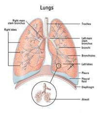 The diaphragm’s job is to help pump the carbon dioxide out of the lungs and pull the oxygen into the lungs. The diaphragm is a sheet of muscles that lies across the bottom of the chest cavity. During the contraction and relaxation of the diaphragm breathing takes place. When the diaphragm contracts, oxygen is pulled into the lungs. When the diaphragm relaxes, carbon dioxide is pumped out of the lungs.
The Cardiovascular System
The primary function of the cardiovascular system is to circulate oxygen throughout our bodies. This system includes the heart, arteries and veins. Arteries carry oxygen-rich blood away from the heart. Veins carry oxygen-poor blood back to your heart.
The heart pumps oxygen into the blood and collects carbon dioxide from it to be expelled through the lungs. The blood starts its journey throughout the body at the right side of the heart. It goes out to the lungs where it exchanges carbon dioxide for oxygen. At the lungs it is the pulmonary artery that brings oxygen-poor blood into the lungs and the pulmonary vein that carries oxygen-rich blood back to the heart. The blood goes back to the left side of the heart and is pumped out to the rest of the body. The main artery from the heart is called the aorta. As the blood travels, it enters smaller and smaller blood vessels, reaching every cell in the body, dropping off nutrients and picking up waste products and carbon dioxide. The blood then starts the trip back in the veins, entering larger and larger ones as it goes, passing through the kidneys and the liver on the way to drop off waste products. The blood eventually arrives back at the right side of the heart to start the trip all over again.
Blood Vessels
The blood vessels carry blood between the heart, different tissues, and organs of the body. There are three types of blood vessels: arteries, veins, and capillaries. These blood vessels have the ability to expand to allow more blood to flow through them. They can also contract to help control the flow of blood. The blood flows through the arteries into the capillaries to the veins. The Veins then return blood to the heart.
Effects of interruption of blood flow to the brain
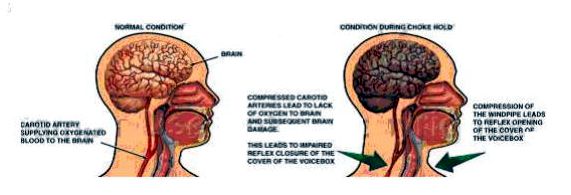
The brain has an absolute dependence on the blood for its immediate supply of oxygen and energy substrates. Interruption of oxygen or substrate supply by a restriction of respiratory or cardiovascular function results in cerebral hypoxy (oxygen deficiency in the brain) creating dizziness, temporary loss of consciousness or possible death.
In shima waza, interruption of oxygen to the brain is usually achieved through applying pressure to various parts of the body, in particular the neck. Therefore a more in-depth review of the neck is required.
The Neck
 The neck contains important communications between the head and the body, including air passages, blood vessels and nerves, and the spinal cord. Many vital structures, such as the visceral column (pharynx, larynx, trachea, and esophagus), major branches of cranial nerves (vagus nerve and the hypoglossal nerve), and major vascular structures (carotid artery, vertebral artery, jugular vein) are compressed into a narrow area, which is engineered for maximal mobility of the head position relative to body.
The sternocleidomastoid, the large muscle located on each side of your neck and used to control movement of the head, forms the boarders of the neck used for anatomical mapping.
Anatomical Mapping of the Neck
For the purpose of shime waza, the neck can be divided into triangles, the two most important being the anterior and posterior cervical triangles.
The anatomic borders of the anterior cervical triangle are the medial portion of the sternocleidomastoid muscle, the lower border of the mandible, and the midline of the neck. Important structures within this triangle are the carotid arteries and jugular veins, esophagus, trachea, larynx, and vagus nerve.
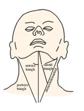 The posterior cervical triangle’s borders are the lateral edge of the sternocleidomastoid muscle, trapezius, and clavicle. Within this region are the subclavian artery and vein, suprascapular artery, and brachial plexus.
Strangulation and Choking
The terms choke and strangulation are often used interchangeably but in fact, refer to two different types of techniques. Strangulation is a form of hypoxia characterized by a temporary cerebral ischemia, i.e. a temporary condition where the oxygen-rich blood flow to the brain is restricted. Choking blocks the internal airway, cutting off the air supply to the lungs.
Categorizing Chokes and Strangulation
Chokes and strangulations can be categorized according to the areas attacked and the resultant outcome it has on the body as follows;
• Vascular Compression
• Tracheal occlusion
• Carotid Sinus Stimulation
Vascular Compression
Vascular compression is a pressing/squeezing of the soft tissues of the neck in and around the area housing the jugular veins and carotid arteries. Vascular compression seeks to induce cerebral hypoxia, a condition in which there is a decrease of oxygen supply to the brain, by direct pressure on the carotid artery and an indirect squeezing’ of the jugular vein by the tensing of sternocleidomastoid muscles (and a host of smaller accessory muscles). Vascular compression requires approximately 4.4 to 11 pounds of pressure with a time interval of compression to loss of consciousness of approximately 10 seconds if both carotid arteries are compressed and a minute if only the jugulars are compressed. The time interval from loss of consciousness to death is said to be in the region of minutes.
Tracheal Occlusion
The larynx is an organ in the neck involved in the control of breathing, protection of the trachea and sound production. The larynx houses the vocal cords and is situated at the point where the upper tract splits into the trachea and the esophagus.
Tracheal occlusion is an application of pressure directly across the front to of the throat, directed towards the trachea or windpipe thus pushing the hyoid bone and tongue are upwards and backwards against the laryngo-pharynx. Tracheal occlusion seeks to prevent the renewal of oxygen of the blood and brings about asphyxia.
Asphyxia, literally translated from the Greek means “absence of pulse”. In its broadest sense it refers to a lack of oxygen that produces a potentially lethal build-up of carbon dioxide. This results in a loss of consciousness and/or death.
Chokes in actual practice are generally effective due to the frightening sensation, discomfort/pain and the resultant panic involved, and not the actual cutoff of oxygen. The effects of tracheal occlusion occur with 33 pounds of pressure.
Carotid Sinus Stimulation
Compression of the neck may also cause stimulation of the carotid sinus with bradycardia (a condition causing a slowing down of the heart rate). The carotid sinus contains baroreceptors (pressure sensors/nerve endings) sensitive to pressure, which when stimulated cause a reflexive slowing of the heart rate, a drop in blood pressure (via dilation of blood vessels), and a subsequent decrease of blood flow to the brain. The symptoms of this sudden decrease of blood to the brain are collectively called syncope, which includes a sudden loss of muscle tone and strength, making it difficult to stand erect, and may cause a loss of consciousness or cardiac arrest. Carotid Sinus Stimulation requires approximately 11 pounds of pressure with a time interval of 10 seconds to loss of consciousness.
Major Targets of the Neck used in Shime Waza
Cartoid Artery
 Carotid artery, originating from Greek word kartoid means amazing deep sleep. It is located in the front of the neck though which blood from the heart goes to the brain.
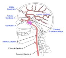
There are 2 carotid arteries — the right and left common carotid arteries — on each side of the neck. Together, the right and left common carotid arteries provide the principal blood supply to the head and neck.
The left common carotid arises directly from the aorta (the huge artery that comes from the heart). The right common carotid artery arises from the brachiocephalic artery, which, in turn, comes off the aorta.
Each of the two common carotid arteries divides to form external and internal carotid arteries. The external carotids are more superficial (closer to the surface) than the internal carotids (which run deep within the neck).
Vagus Nerve
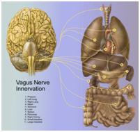 Vagus nerve, meaning, “wandering”, is the tenth cranial nerve. It originates in the medulla oblongata, a part of the brain stem, and wanders all the way down from the brainstem to the colon.
The vagus nerve runs to the heart parallel to the common carotid artery. Its function is to regulate heart rate, which affects blood pressure.
Interruption of the vagus nerve causes a decreasing of the heart rate, lowers blood pressure and hence lowers the volume of oxygenated blood to the brain.
Jugular Vein
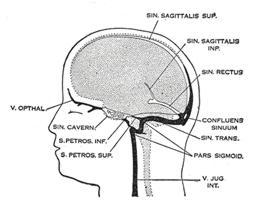 Jugular vein, derived from the Latin “jugulum” meaning throat or collarbone, is located in the neck and drains blood from the head, brain, face and neck and conveys it toward the heart.
The external jugular vein collects most of the blood from the outside of the skull and the deep parts of the face. It lies outside the sternocleidomastoid muscle, passes down the neck and joins the subclavian vein.
The internal jugular vein collects blood from the brain, the outside of the face and the neck. It runs down the inside of the neck outside the internal and common carotid arteries and unites with the subclavian vein to the innominate vein.
Improvised Weapons
In the execution of shime waza, the arsenal of possible body parts used in the performance of a choke or strangle includes the fingers, knuckles, wrists, hands, forearms, foot, shin, knees and legs. Other possible non-body implements used could be clothing (e.g., jacket lapel) or implements (use of a belt or a club against the throat).
Entries
In applying chokes in which you wrap the neck, there are two basic methods of entry: a) circle the neck raking with your thumb knuckles; b) Execute a strike which places your attack point at the target to be choked.
If an attacker drops their chin to protect the neck, press sensitive points to move their head;
• TW-17 (behind the ear)
• ST-4 (jaw)
• GB-1 or TW-23 (temple)
• Eyes and ST-2 (cheek)
Eliminating Slack
When applying shime waza, it is very important to eliminate an slack. The effectiveness of shime waza is reduced when there is any slack or movement in the hold and will encourage escape and counters. During a choke make sure the part of the body you are applying the choke with is wrapped tight against the surface you are attacking.
To restrict movement:
1. Use the lapel
2. Use the wall
3. Use the ground
4. your arm
5. your head
6. the attackers weight
7. your weight
Practical Concerns
Effects and Possible Consequences of Shime Waza
The physiological responses to the three categories of chokes and strangulation is a decrease in motor coordination, loss of consciousness, or if sustained for a sufficient interval (minutes) death. The application of chokes and strangulation can also damage the internal structures of the neck like the hyoid bone, larynx, trachea, etc. or cause brain damage. Needless to say, applying these techniques carry some risk and should be applied only in life threatening situations.
The law in many countries view choking and strangulation techniques as excessive use of force and in the event death occurs the person applying the technique could be convicted of manslaughter because having to hold pressure for two solid minutes is good evidence of intent.
Basic Techniques of Resuscitation
Be prepared to resuscitate your opponent should it be appropriate. In a very few cases, the person does not regain consciousness and emergency first aid must be administered.
Positioning: An unconscious person should be rolled on to their side while protecting the head and letting it rest on their extended arm in the recovery (lateral recumbent) position. In this position, the airway remains open and is not blocked by a relaxed tongue. Fluids such as saliva and maybe even vomit are also allowed to flow out of the mouth and not into the airway.
Unresponsiveness: Try to awaken the person with vocal stimuli (shout and call out to the athlete). Try to awaken the person with pain stimuli (e.g. triceps pinch, slap on the back or sternal rub.)
Airway: Open and maintain an airway for the person.
Breathing: Check for breathing … Look at the chest rise, listen for air exchange, and feel for a breath. Initiate 2 slow breaths if there is no breathing,
Circulation: Check for the presence of a carotid pulse. If absent, commence chest compressions and artificial respiration.
If the person does not regain consciousness within 20 seconds alert medical services.
Limitations of Shime Waza
In a hostile situation, the situation is dynamic and can change very quickly. As you react to the initial attack, the attacker is probably going to change his attack in reaction to your reaction or resist any technique applied to control him. Therefore, your arsenal of techniques must be able to react to quickly changing situations and you must arm yourself with the most efficient tool. While shime waza represent a very effective tool for self-defense, we must bear in mind that they can also fail for many reasons. Reasons include the improper application of your technique, resistance and counters by your attacker, the changing situation.
Conclusion
It is dangerous to imply that there is a single solution to all threatening situations. There is, in fact, a range of responses available to you. The situation and circumstances will dictate which of them is most appropriate.
Many believe attacking your opponent’s air/blood supply, through the application of shime waza, is one of the most effective and most humane ways of defending oneself. Others feel that it is best not to attempt shime waza in a self-defense situation because the ultimate outcome of shime waza could be death to your attacker if applied by someone not properly trained. If your attacker dies you may then be considered the aggressor and face possible criminal prosecution.
Whichever side of the fence you are on with regard to this debate, it is essential to understand how to respond to a violent confrontation and the potential consequences of your actions. Unfortunately, during the haste of a self-defense situation, we must make some judgment whether an attacker is determined on doing you bodily harm and a quick decision on the appropriate coarse of action (technique) to take for your survival.
References:
Koiwai EK. Journal of Forensic Sciences – Deaths Allegedly Caused by the Use of ‘Choke Holds’ (Shime-Waza)
Arsenault, Alan, D. Chin Na in Ground Fighting: Principles, Theory and Submission Holds for all Martial styles. Boston, YMAA Publications, 2003.
Reay Donald, T. and J.W. Eisele, “Dearth From Law Enforcement Neck Holds.” The American Journal of Forensic Medicine and Pathology. Vol. 3, No. 3: 253-258, 1982.
Siddle, Bruce K. Pressure Point and Control Tactics: Defensive Tactics Instructor Manual. Millstadt: PPCT Management Systems Inc., 1999.


